Ultrafast 3D histological imaging based on a minutes-time scale tissue clearing and multidirectional selective plane illumination microscopy
Jiajia Chen, Zhenhong Du, Chenhao Xu, Xiao Xiao, Wei Gong, and
Ke Si
Optics Letters, 2022
, 47, 17, 4331–4334
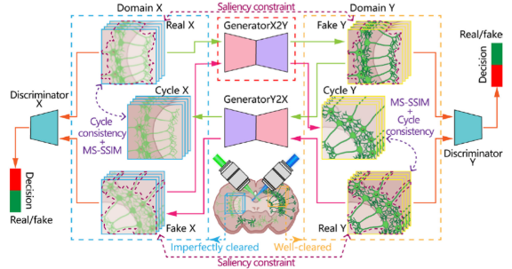
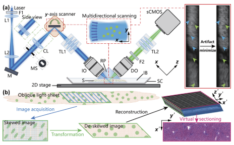
Conventional histopathological examinations are time-consuming and labor-intensive, and are insufficient to depict 3D pathological features intuitively. Here we report an ultrafast 3D histological imaging scheme based on optimized selective plane illumination microscopy (mSPIM), a minutes-time scale clearing method (FOCM), and a deep learning-based image enhancement algorithm (SRACNet) to realize histological preparation and imaging of clinical tissues. Our scheme enables 1-minute clearing and fast imaging (up to 900 mm2/min) of 200 \textmum-thick mouse kidney slices at micron-level resolution. With hematoxylin and eosin analog, we demonstrated the detailed 3D morphological connections between glomeruli and the surrounding tubules, which is difficult to identify in conventional 2D histology. Further, by the preliminary verification on human kidney tissues, this study will provide new, to the best of our knowledge, feasible histological solutions and inspirations in future 3D digital pathology. \copyright 2022 Optical Society of America